The first FBDD-related meeting of 2016 has already come and gone, but there are still plenty of events ahead. Below are several updates as well as a new listing.
April 20-21: CHI’s Eleventh Annual Fragment-Based Drug Discovery, the longest-running fragment event, will be held in San Diego. You can read impressions of last year's meeting here, here, and here; the 2014 meeting here and here; the 2013 meeting here and here; the 2012 meeting here; the 2011 meeting here; and 2010 here. Also as part of this event, Ben Davis and I will be teaching a short course on FBDD over dinner on April 20.
May 24: Development of Novel Therapies through Fragment Based Drug Discovery will be held in Houston, Texas. Despite being only one day, it looks like a great lineup of speakers, so check it out!
June 7-10: Although not strictly fragment-focused, the third NovAliX Conference on Biophysics in Drug Discovery is likely to have lots of relevant talks, and is a good excuse to get to Strasbourg, France. You can read Teddy's impressions of the 2013 event here, here, and here.
July 12-15: FBDD Downunder 2016 will be held at Monash University in Melbourne. This is only the second such event; the first was lots of fun and even resulted in a special issue of the Aust. J. Chem., so definitely check this out if you can.
October 9-12: FBLD 2016 will be held in Boston, MA. This marks the sixth in an illustrious series of conferences organized by scientists for scientists, the last of which was in Basel in 2014. Surprisingly, this also seems to be the first dedicated fragment conference in Boston. You can read impressions of FBLD 2012, FBLD 2010, and FBLD 2009. Early-bird registration is now open!
November 7-9: Finally, the OMICS Group is holding their second Drug Discovery & Designing in Istanbul, Turkey, with a track on FBDD.
Know of anything else? Add it to the comments or let us know!
29 February 2016
22 February 2016
Fragments vs PRMT6
Epigenetics involves turning
genes on or off without changing their sequence. This often relies on modifications
to proteins or DNA that are recognized by other proteins. As Teddy pithily
observed, this is a big field. However, in the realm of fragments, most of the
attention has been on bromodomains; other classes of proteins, such as
methyltransferases, have been largely neglected. A new paper in J. Med. Chem. by Masoud Vedadi and
Matthieu Schapira and collaborators at the University of Toronto and Bayer
suggests fragments are promising for these targets as well.
The researchers were specifically
interested in protein arginine methyltransferases (PRMTs), which transfer a
methyl group to one of the terminal side chain nitrogen atoms on specific
arginine residues. PRMT6 in particular targets histone proteins to modulate
transcription and has been implicated in cancer as well as neurodegenerative
diseases. A few potent inhibitors have previously been reported for PRMTs, and
the team started by deconstructing them to hunt for active fragments.
Ligand deconstruction involves
chopping a known ligand into fragments to see whether any of these pieces will
still bind. In this particular case, EPZ020411 had previously been
characterized crystallographically bound to PRMT6 with the basic
amine-containing “tail” in the substrate arginine-binding groove. Testing this
fragment 6 by itself revealed a low micromolar inhibitor with a ridiculously
high ligand efficiency.

Thus encouraged, the researchers
ran a functional screen of 2040 diverse fragments (about half from Maybridge)
at 1 mM concentration and retested hits at 0.5 mM. About half the resulting
hits were false positives or other uglies, leaving the researchers with 14
fragments with IC50 values from 0.3 – 400 µM. As might be expected given the cationic nature of the substrate, 12 of these have basic
nitrogens.
Compound 7 was particularly
interesting: at 300 nM this is one potent fragment! ITC revealed a dissociation
constant of 970 nM, with a favorable enthalpy and unfavorable entropy of binding. It did hit other PRMTs, but was
remarkably selective against a panel of 25 other human methyltransferases.
The researchers also determined
the crystal structure of compound 7 bound to PRMT6, which revealed it binding,
as expected, in the arginine site, making hydrogen bonds with a conserved
catalytic glutamic acid. Weirdly though, it seems to be a noncompetitive
inhibitor: increasing concentrations of substrate peptide or cofactor had no
effect on inhibition. The team speculates that the noncompetitive behavior
could be because the substrate makes strong interactions with the protein
outside the arginine-binding site. Nonetheless, the fragment did inhibit PRMT6
activity in a cell assay with IC50 = 21 µM.
Overall then it seems that the
PRMTs are amenable to FBLD. They are interesting drug targets, and at the very
least having more probes will help to unravel the biology.
15 February 2016
Selectivity in STD
Among NMR-based fragment
screening methods, saturation transfer difference (STD) came in as most popular
in a recent poll. The technique is very sensitive and thus able to identify
weak fragments. Unfortunately, it’s a bit too sensitive; hit rates of >30%
are not uncommon. Many of these hits interact non-specifically with the
protein. These can be weeded out using orthogonal screening methods or
competition assays, but it would be nice to make STD itself more
discriminating. In a paper published late last year in J. Med. Chem., Olivier Cala and Isabelle Krimm describe how to do
just this.
In an STD experiment, the protein
target is irradiated and transfers some of its magnetization to bound ligands.
When these dissociate they retain some of the magnetization, and so the NMR
signals of the fragments decrease. The problem is that lots of fragments interact
non-specifically with proteins. For example, if a lipophilic fragment dances
across greasy patches on a protein surface to escape from water without making
any specific interactions, it will still get magnetized. Can such signals be
distinguished from fragments that bind in a single, well-defined manner?
Within a ligand that binds to a
protein, a proton that binds closer to the protein will show a stronger STD
effect than one that is exposed to solvent. This is in fact the basis for STD “epitope mapping”, which allows one to roughly model how a ligand binds to a protein –
or at least which parts of a ligand are closest to the protein. In the new
paper, the researchers argue that simply observing differences in the STD
effect between different protons in a ligand can distinguish whether or not
that ligand is binding in a single binding mode.
Several examples support this
assertion. For one protein, all the fragments that showed significant epitope
mapping could be competed with a known reference molecule, suggesting binding
to a specific site; this was less often the case for fragments that did not
show epitope mapping. In another example, the privileged fragment 7-azaindole
was found to bind to two different proteins with different epitope maps,
suggesting different (but specific) binding modes for each protein. The
technique also seems fairly robust to the affinity of the fragments (KD
50 µM to > 1 mM), the details of the NMR experiment (saturation time from
0.5 to 4 seconds), and ligand/protein ratios between 66 to 1 and 400 to 1.
As the researchers note, there
are caveats. For example, if a fragment can bind in two different but
nonetheless specific binding modes, it may show uniform STD effects and will be
a false negative. Nonetheless, comparing STD effects across a ligand does seem
a worthwhile exercise. Not only could it help prioritize fragments, it could
also reveal which protons are further from the protein, and so suggest growth
vectors.
08 February 2016
Dihydroisoquinolones as fragments
It’s a common problem: you find a
fragment that binds to your target and want to grow it to improve affinity. A search
for commercial analogs comes up empty, so you look into modifying the hit, only
to discover that you’ve got a six-step synthesis on your hands. Or worse;
perhaps there is no precedent at all. The chemical literature is replete with
total syntheses of complicated natural products, but seemingly simple fragments
are often not well-represented. Last year, researchers from Astex exhorted
chemists to develop synthetic routes for attractive fragments, and in a recent paper in Org. Biomol. Chem. David
Rees and colleagues take up their own challenge in the case of dihydroisoquinolones.
Dihydroisoquinolone itself is a
nifty little fragment. It has just 11 atoms, cLogP = 1.0, and its solubility is > 5 mM in aqueous buffer. Its cis-amide
moiety can serve as a hydrogen bond donor and acceptor, and the adjacent phenyl
ring provides a bit of grease for interacting with hydrophobic protein residues.

The researchers built on existing
methodology using a rhodium catalyst to introduce polar groups (such as
hydroxymethyl and dimethylamino) at the R position. Depending on the nature of
the R group, regioisomers in which the substituent ends up at the 4-position
could sometimes also be isolated.
The methodology is robust and tolerates
air, moisture, and various substituents. The alkene starting material is easy
to come by, and the aromatic starting material is easy to make. By varying this,
the researchers could generate 6- or 7- substituted dihydroisoquinolones,
though 5- and 8- substituted versions seem harder to access. The team was also able
to use other aromatics as starting materials, including thiophene, thiazole,
and pyridine.
Thus, if dihydroisoquinolone
comes up as a hit, this paper will allow you to quickly explore most of the
vectors. So how often does this fragment show up? It is not clear why some
fragments, such as 7-azaindole and 4-bromopyrazole, show up again and again,
while others languish so lazily in the library that they might as well not even
be there. We’ve highlighted at least one case where a dihydroisoquinolone was a useful hit.
Practical Fragments would love to know your experience. Do you have
dihydroisoquinolones in your library? How often do they show up as hits? And
what other fragments do you find that are in need of better synthetic routes
for further exploration?
01 February 2016
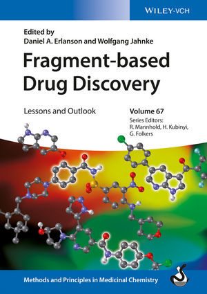
Fragment-Based Drug Discovery: Lessons and Outlook
In 2006, Wolfgang Jahnke and I co-edited the very first book
on fragment-based drug discovery. Half a dozen books have followed, most of
which have been reviewed at Practical
Fragments (see right-hand column). These are now joined by a new book edited
by Wolfgang and me in Wiley’s Methods and
Principles in Medicinal Chemistry series.
At 500 pages and 19 chapters, this is the most extensive
treatment since the Methods in Enzymology
volume five years ago. In the interest of space I can’t write more than a
sentence or two about each chapter, but I would like to thank all the
contributors. Although I’m undoubtedly biased, I believe this work will set the
standard for years to come.
The book is divided into three sections, starting with The Concept of FBDD. Rod Hubbard
(Vernalis and University of York) opens with a chapter on the role of FBDD in
lead-finding, which provides an introduction, historical overview, and summary
of current thinking and future challenges. One particularly interesting section
compares the contents of the 2006 book with the state of the art today,
highlighting the fact that many of the basic techniques were already in place a
decade ago, but the number of success stories has increased dramatically.
Chapter 2, by Glyn Williams and colleagues at Astex,
discusses how to choose targets for FBDD, including concepts such as
ligandability. Key principles are nicely illustrated with several important
targets including the IAPs and HCV-NS3.
The last two chapters in this section focus more on numbers.
Chapter 3, by Jean-Louis Reymond and colleagues at the University of Berne, covers the computational enumeration of chemical space, with a special emphasis
on the contents and uses of their GDB-17 set of the 166 billion possible molecules
with up to 17 non-hydrogen atoms. And chapter 4, by György Ferenczy and György
Keseű at the Hungarian National Academy of Sciences, provides an overview of
various metrics (such as ligand efficiency and LELP) and how these can be
useful for fragment optimization.
The next nine chapters comprise the longest sub-section of
the book, Methods and approaches for
FBDD. To start screening fragments, you need a library, and designing one
is the subject of chapter 5, by Martin Drysdale and colleagues at the Beatson
Institute. This chapter also touches on concepts such as molecular complexity
and “three-dimensional” fragments.
Screening techniques are best used in combination, and in
chapter 6 Ben Davis (Vernalis) and Tony Giannetti (Google[x]) describe the
synthesis of results from SPR, NMR, X-ray, ITC, functional screens, and other
techniques to overcome challenges in several discovery programs. They emphasize
that universal agreement among different methods is not always necessary, but
carefully analyzing discrepancies can reveal unexpected problems with the
screening conditions, target, or hits.
Differential scanning fluorimetry (DSF) – or thermal shift
(TS) – is perhaps the most controversial screening method, and in chapter 7
Chris Abell and colleagues at the University of Cambridge cover this approach
in depth. The chapter starts with a thermodynamically detailed yet nonetheless
lucid discussion of the theory behind DSF, including the interpretation of
negative thermal shifts. The chapter also includes plenty of practical advice
and case studies, some of which we’ve covered briefly (for example here and
here).
Chapter 8, by Sten Ohlson and Minh-Dao Duong-Thi at Nanyang
Technological University, covers three emerging fragment screening
technologies: WAC, native MS, and MST. And Chapter 9, by Sandor Vajda (Boston
University) and collaborators, does an excellent job of summarizing
computational approaches.
As others have noted, some of the biggest challenges are not
technical but organizational, and in chapter 10 Michelle Arkin and colleagues
at UCSF describe how to make FBDD work in academia. The chapter also includes
some interesting polling data, concise but cogent summaries of fragment-finding
techniques, and case studies on p97 and caspase-6. And in chapter 11, Jim Wells
and colleagues – also at UCSF – describe using Tethering to find allosteric sites in proteins.
One area that has grown dramatically since 2006 is the use
of FBDD in complex systems (such as membrane proteins), the subject of a
chapter by Miles Congreve and John Christopher at Heptares. Chapter 12 also
includes successful case studies, some of which we’ve covered. But finding
fragments against these targets is still not easy, as illustrated in the final
figure: of 18 fragment hits on 15 targets, almost all have ligand efficiency
values > 0.3 kcal/mol per atom, and most of them are relatively potent, with
affinities in the mid-micromolar range or better. While everyone wants to find
strong binders from the start, such numbers suggest many weak-binding hits are overlooked.
Chapter 13, by Jörg Rademann and colleagues at Freie
Universität Berlin, covers protein-templated fragment ligation methods, both
reversible and irreversible. The chapter is wide-ranging and includes methods
such as dynamic libraries and various types of “Click” chemistries.
The last section of the book, which was mostly absent a
decade ago, is entitled Successes from
FBDD. This starts with a chapter by Daniel Wyss, Andrew Stamford, and
colleagues from Merck on BACE inhibitors. As we’ve noted, fragments have had a
major role in most of the BACE inhibitors to enter the clinic, with phase III
results from Merck’s verubecestat expected next year.
Epigenetics has also been strongly influenced by fragments,
and in chapter 15 Aman Iqbal (Proteorex) and Peter Brown (Structural Genomics
Consortium) survey the field, with case studies on several proteins that modulate
epigenetic marks. These include BRD4, ATAD2, BAZ2B, SIRT2, and others.
One of the original selling points of fragment-based methods
is the ability to go after difficult targets such as protein-protein
interactions, and this is the subject of chapter 16, by Feng Wang and Stephen
Fesik (Vanderbilt University). In addition to general guidelines, the
researchers describe a number of case studies, including RPA, MCL-1, and K-Ras.
Some enzymes can be just as difficult as protein-protein
interactions, and in chapter 17 Alexander Breeze (University of Leeds) and
former AstraZeneca colleagues describe programs to find inhibitors of LDHA (see
here and here). They also discuss how some previously reported inhibitors
turned out to be artifacts.
More than two dozen kinase inhibitors have been approved by
the US FDA, including the first drug derived from FBDD. In chapter 18,
Gordon Saxty (Fidelta) surveys a number of kinase programs, including most of
the fragment-derived inhibitors in clinical trials.
And finally, in chapter 19 Simon Rüdisser and colleagues from
Novartis present an extensive discussion of renin, with special attention to
their campaign, which involved a combination of HTS and fragment-based
approaches.
Subscribe to:
Posts (Atom)

